sc-RNA-Seq Analysis: The Immune Changes Induced by Intratumoral Hapten Plus Chemotherapy Drugs of Endometrial Cancer
2 Department of Oncology, Jinan Baofa Cancer Hospital, Jinan, Shandong Province, China
3 Department of Oncology, Beijing Baofa Cancer Hospital, Beijing, China
4 Department of Oncology, TaiMei Baofa Cancer Hospital, Dongping, Shandong, China
5 Department of Oncology, Immune Oncology System, San Diego, California, USA
Peicheng Zhang, Department of Oncology, Royallee Cancer Hospital, Guangzhou, China, Tel: +86 105 615 2159, Email: bfyuchina@126.com
Received: 29-Jun-2023, Manuscript No. jbclinphar-23-104208; Editor assigned: 03-Jul-2023, Pre QC No. jbclinphar-23-104208; Reviewed: 01-Jul-2023 QC No. jbclinphar-23-104208; Revised: 24-Jul-2023, Manuscript No. jbclinphar-23-104208; Published: 31-Jul-2023
Citation: Yu B, Zheng G, Jing P, et al. sc-RNA-Seq Analysis: Intratumoral Hapten Inducing Immune Changes and Chemotherapeutic Drugs of Endometreal Cancer. J Basic Clin Pharma. 2023,14(S1):4-10
This open-access article is distributed under the terms of the Creative Commons Attribution Non-Commercial License (CC BY-NC) (http://creativecommons.org/licenses/by-nc/4.0/), which permits reuse, distribution and reproduction of the article, provided that the original work is properly cited and the reuse is restricted to noncommercial purposes. For commercial reuse, contact reprints@jbclinpharm.org
Abstract
Study of intratumoral hapten plus chemotherapy drugs for endometrial cancer, It was found that hapten enhanced intratumoral chemotherapy treatment of endometrial cancer induced acute immune response followed the hysterectomy. The results of Gene Set Variation Analysis (GSVA) analysis showed enrichment after treatment: non-classical NF-kB activation, and the non-classical Nuclear Factor Kappa Light chain enhancer of activated B cells (NF-κB) pathway was responsible for the development of multilayer immune cell;there were high expression of T cell toxicity (CCL3, CLL4, GZMB, GZLY) and inflammation (CCL5, NGK7) genes in the intermediate stage, Conventional Dendritic Cells (cDCs), the macrophage was increased in the microenvironment after treatment, T cells was increased after treatment as important factor to kill tumors in the microenvironment. The intercellular communication in Major Histocompatibility Complex (MHC I) pathway of concern showed that Antigen Presenting Cells (APC) and T cells in MHC I pathway had obvious communication before and after treatment. Hapten with chemotherapy treatment resulted in significantly clinical benefit, one local therapy can kill local endometrial cancer also induce immune response to fight cancer with or without hysterectomy and control expression of several genes related with endometrial cancer.
Our study provides evidence that hapten mediated local chemotherapy is safe and effective method while it induces a systematic immunity against cancer by initiating immune response from the endometrial cancer to achieve desirable clinical outcome in order to prevention of tumor cell metastasis with or without hysterectomy, in fact, it bring the immunotherapy advance ahead any treatment of cancer, cleverly integrated into existing therapies and make those quite effective in prolong patient’s life
Keywords
Hapten; Chemotherapy Drugs; Endometrial Cancer; Hysterectomy
Introduction
About 142,000 women worldwide develop new cases of endometrial cancer, and about 42,000 women die from this cancer. The typical age of endometrial cancer - the incidence is highest around the seventh decade of life, and most cases are diagnosed after menopause. Most women with endometrial cancer are in the early stages when they develop the disease, with an overall 5-year survival rate is about 80 percent. The cornerstone of the treatment of endometrial cancer is surgery, which is not only important for staging, but also suitable for adjuvant treatment modalities that benefit high-risk patients [1]. Hysterectomy and bilateral salpingo-oophorectomy with or without lymphadenectomy is painful for women interested in future fertility [2]. Postoperative adjuvant treatment of endometrial cancer and advanced disease, with emphasis on chemotherapy, radiation, and a combination of the two. These treatments are tailored to the risk of clinical recurrence [3].
Immunotherapy checkpoint inhibitors have become a new model for the treatment of multiple tumors, and endometrial cancer is no exception. Most recently pembrolizumab /lenvatinib for all patients with endometrial cancer. Endometrial cancer is a heterogeneous disease with different molecular subtypes and different prognoses. Differences between molecular subpopulations regarding antigenicity and immunogenicity should be relevant for the development of more targeted immunotherapy approaches [4].
There is few research how to treat endometrial cancer with new option since it is a rare disease. Ultra-Minimum Incision Personalized Intratumoral Chemoimmunotherapy (UMIPIC) is used treated most solid tumors in past few years, it need to try for care endometrial cancer as a new tool for treating cancer [5-7]. The recent progress of single-cell RNA sequencing (scRNA-seq) allows scientists to explore the genetic and functional heterogeneity of cellular complexes at the molecular level [8]. Since single-cell transcription profiles provide a suitable alternative method, direct comparisons between the same cell types can now be made before and after treatment. However, the current single-cell studies of endometrial cancer mostly focus on the Tumor Micro Environment (TME) [9]. There is still a lack of comparative studies of immune reaction changes at the single-cell level for male breast cancer before and after hapten enhanced UMIPIC.
In the current study, we used scRNA-seq to investigate and compare the changes in myeloid cells, stromal cells, T cells, plasma cells, B cells, platelets, erythrocytes, epithelial cells in endometrial cancer before and after treatments of UMIPIC. We showed that the immune cells were awakened to aid cancer immunotherapy like Programmed Death Ligand (PD)1 or PD-l1 to achieve immune response and it may be useful for prevent tumor metastasis or recovery [6,7]. Our study may provide a detailed understanding of immune cell awakened at the molecular base in fighting cancer cells that facilitates the development of a more reliable, hapten enhanced sensitization immunotherapy for treatment of patients with endometrial cancer with or without hysterectomy.
Materials and Methods
Clinical specimens
Patient was diagnosed as endometrial cancer, consistent with MRI findings of the tumor (endometrial cancer, stage Ib), left uterine myoma. the pathological results showed that:(uterine) endometrial glandular hyperplasia was dense, the interstitium was less, and the local interstitium showed multiple foam cell infiltration, immunohistochemistry of Estrogen Receptor(ER) Progesterone Receptor (PR), Phosphatase and Tensin homolog (PTEN), CK/5, P53, KI-67, CD10 and mismatch repair protein (MLH1MSH2MSH6PMS2) is further confirmed diagnosis of endometrioid adenocarcinoma. Immunohistochemistry:(uterine cavity) combined morphology and immunohistochemistry suggested atypical endometrial hyperplasia, focal carcinomatous transformation into endometrioid adenocarcinoma, grade I. No. 3 Immunohistochemistry: ER+(range about 90%, strength: strong), PR:+(range about 90%; Intensity: strong), PTEN:(-), CK5/6:(individual +), P53:about 60% positive cells, Ki-67: about 30% positive cells, CD10: a very small amount (+), mismatch repair protein immunohistochemical results has suggested tumor satellite instability (dMMR); MLHI+, PMS2-, MSH2+, MSH6+. Intrauterine drug infusion therapy was performed with 10 ml drugs combination solution to whole uterine cavity that contained 1.00 mg/ml Adriamycin (Adr), 0.80 mg/ml of Cytarabine (Ara-C), 20.0 mg/ml of H2O2 and 144 mg/ml of penicillin as the hapten and one week later, transabdominal extrascial hysterectomy plus double adnexectomy was performed under tracheal intubation and general anesthesia. The operation was successful. The excised specimens were routinely sent for pathological examination after being examined by a group of the patient and her family and confirm the effective of treatment. One piece of excised specimen took as treated samples for scRNA-Seq analysis.
Tissue disassociation and cells collection
After small endometrial cancer tissues before and after treatment were collected, the fresh tissue samples were immediately stored in the sCelLiVE® Tissue Preservation Solution (Singleron) on ice. The tissues were cut into small tissue pieces and were transferred to a 15-ml centrifuge tube, followed by digestion using sCelLiVE® Tissue Dissociation Solution (Singleron) at 37°C for 15 min with shaking. The samples were then filtered with 40 µm sterile strainers, and centrifuged at 1,000 rpm at 4°C for 5 min. Next, 2 ml GEXSCOPE® Red Blood Cell Lysis Buffffer (RCLB, Singleron) was added to lyse the red blood cells for 10 min. Finally, the single cell suspension was collected after re-suspension with Phosphate Buffered Saline (PBS), and trypan blue (Sigma) staining was used to calculate cell activity and cell count under a microscope.
Single-cell RNA sequencing
Single-cell suspensions (1~3×10⁵ cells/mL) in PBS (HyClone) were loaded onto microwell chip using the Singleron Matrix® Single Cell Processing System. Briefly, the scRNA-seq library was constructed using the GEXSCOPE® Single Cell RNA Library Kits (Singleron). The library was lastly sequenced with 150bp was diluted to 4nM and paired-end reads on the IlluminaHiSeq X platform following an established protocol [10]. Sequencing data processing and quality control was performed as described in previous publications [11].
Differentially Expressed Genes (DEGs) analysis
To identify Differentially Expressed Genes (DEGs), genes expressed in more than 10% of the cells were selected in both of the compared groups of cells and with an average log (fold changes) value greater than 1 as DEGs. Adjusted p value was calculated by the benjamini-hochberg correction. The P value of 0.05 was used as the criterion to assess the statistical significance.
Cell type annotation
The cell type identity of each cluster was determined with the expression of canonical markers found in the DEGs using SynEcoSys database (Singleron Biotechnologies). Heatmaps/dot plots/violin plots displaying the expression of markers used to identify each cell type were generated by the scanpy built-in functions and ggplot2.
Single-Cell Copy Number Variation (CNV) analysis
The InferCNV package was used to detect the CNAs in malignant cells. Non-malignant cells (T and NK cells) were used as control references to estimate the CNVs of malignant cells. Genes expressed in more than 20 cells were sorted based on their loci on each chromosome. The relative expression values were centered to 1, using 1.5 standard deviation from the residual-normalized expression values as the ceiling. A slide window size of 101 genes was used to smoothen the relative expression on each chromosome, to remove the effect of gene-specific expression.
Pathway enrichment analysis
To investigate the potential functions of DEGs between clusters, the Gene Ontology (GO) and Kyoto Encyclopedia of Genes and Genomes (KEGG) analysis were used with the “clusterProfiler” R package 3.16.1. [9]. The GO gene sets including Molecular Function (MF), Biological Process (BP), and Cellular Component (CC) categories were used as references. Pathways with the adjusted p value less than 0.05 were considered as significantly enriched.
Trajectory analysis
Monocle 2 algorithm was used for pseudo-time trajectory analysis, and the dimensionality reduction method used was DDRTree [12].
Intra-Tumoral Heterogeneity (ITH) score calculation
The Intra-Tumoral Heterogeneity (ITH) score was defined as the average Euclidean distance between the individual cells and all other cells, in terms of the first 20 principal components derived from the normalized expression levels of highly variable genes. The highly variable gene was identified using the “FindVariableGenes” function in the Seurat package, with default parameters.
Cell-Cell interaction analysis (CellPhoneDB)
Cell-Cell Interaction (CCI) between B cells, Epithelial cells, Fibroblasts, Mononuclear phagocytes, Mast cells, Neutrophils, T and NK cells were predicted based on known ligand–receptor pairs by Cellphone DB v2.1.0 [13-15].
Results
Clinical benefit characteristics
After treatment UMIPIC to endometrial cancer, post treatment pathological diagnosis is endometrial cancer stage IA. Patient feel better after hysterectomy, every examined
Landscape of single cell transcriptome sequencing before and after lung cancer treatment
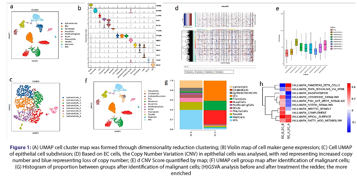
There was a total of 11,729 cells and 12 cell types were constructed for 2 samples. The proportion of stromal cells increased after treatment, which may be due to the thinning of the endometrium after treatment leading to the internal tissue being taken during the sampling process. Therefore, the proportion of cells in the immune microenvironment was explained by subdivision annotation. The epithelial cells obtained at the annotation stage of the large class were further subdivided and 8 epithelial subgroups were obtained. IinferCNV analysis was performed on the epithelial subgroups to determine malignant cells. Clusters 1,6,8 were defined as epithelial cells and cluster 8 as ciliated cells through inferCNV analysis. The remainder are defined as tumor cells.
After treatment, the number of tumor cells was significantly reduced, indicating the possible effectiveness of the treatment. The results of GSVA analysis showed enrichment after treatment: non-classical NF-kB activation, and the non-classical NF-κB pathway was responsible for the development of multilayer immune cells (Figures 1A-1F).
Figure 1: (A) UMAP cell cluster map was formed through dimensionality reduction clustering; (B) Violin map of cell maker gene expression; (C) Cell UMAP of epithelial cell subdivision; (D) Based on EC cells, the Copy Number Variation (CNV) in epithelial cells was analysed, with red representing increased copy number and blue representing loss of copy number; (E) d CNV Score quantified by map; (F) UMAP cell group map after identification of malignant cells; (G) Histogram of proportion between groups after identification of malignant cells; (H)GSVA analysis before and after treatment the redder, the more enriched
Investigate the changes and functional mechanisms of T cells before and after treatment
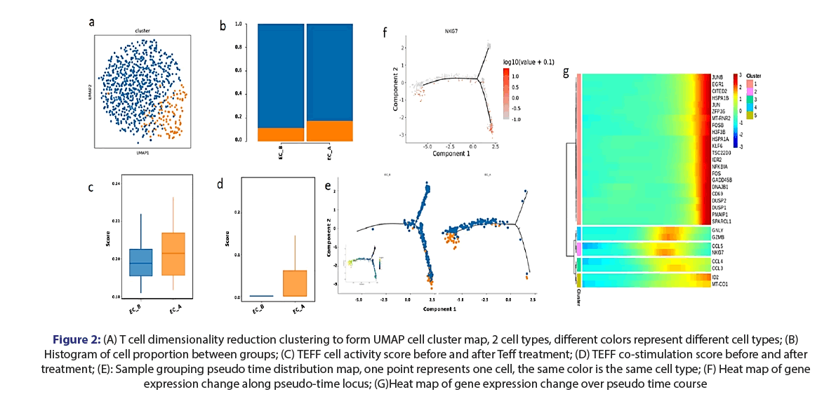
After subdivision annotation of TandNK, two cell types were obtained. After treatment, the number of naiveT cells decreased and the proportion of Teff increased, suggesting that naiveT cells recognize MHC-antigenic peptides presented by Dendritic Cells (DC), monocytes or B cells further differentiate into various T cell subsets. Precisely, the proportion of Teff increased, indicating that cytokines in the microenvironment has promoted its differentiation into Teff, which was rapidly activated, prolificated, migrated to the site of inflammation after being stimulated by antigen, and released immunoactive substances for immune response.
In the absence of the co-stimulatory signals provided by co-stimulatory molecules, T cells cannot be activated, and are in a state of energy, or even T cell apoptosis. Both steps related to T cell activation were effectively improved after treatment. The results showed that hapten therapy could regulate T cell activation to some extent. The results of trajectory analysis showed that there were differences in T cell differentiation before and after treatment: shows the entire trajectory before and after treatment; the cells in the samples before treatment are located in the middle and early stage of the trajectory, while the cells in the samples after treatment were at an advanced stage of the trajectory; before treatment, navieT cells at the beginning of differentiation were mainly present in the samples. After treatment, the number of navieT cells in the sample decreased, and the only remaining navieT cells were those in the TEFF transition stage (in the late stage of differentiation) (Figure 2A-2G).
Figure 2: (A) T cell dimensionality reduction clustering to form UMAP cell cluster map, 2 cell types, different colors represent different cell types; (B) Histogram of cell proportion between groups; (C) TEFF cell activity score before and after Teff treatment; (D) TEFF co-stimulation score before and after treatment; (E): Sample grouping pseudo time distribution map, one point represents one cell, the same color is the same cell type; (F) Heat map of gene expression change along pseudo-time locus; (G)Heat map of gene expression change over pseudo time course
Functional transformation of DC cells in myeloid immune cells before and after treatment
MP cells were subdivided and annotated, and 3 middle myeloid cell types were obtained. The proportion of cDCs increased and DC cells increased as the main APC cells in the microenvironment after treatment, suggesting that the antigen presenting ability of DC cells also increased after treatment, promoting T cells to kill tumors. Although the proportion of macrophages did not change significantly.
After treatment, the proportion of DC cells increased, so the functional transformation of DC before and after treatment was further studied. The improvement of antigen presenting function of DC cells in tissues after hapten treatment. B7-1 (CD80) and B7-2 (CD86) co-stimulatory factors, inter-group gene expression analysis showed that co-stimulatory analysis was also increased in DC after treatment. The significant enrichment of MHC-related pathways in DC cells also suggests that T cell activation is driven by MHC signals provided by DC cells. The co-stimulating molecule CD86 during T cell differentiation was also expressed in the T differentiation locus (Figure 3A-3F).
Figure 3: (A) Color UMAP by cell type; (B) Single sample cell composition histogram; (C) Go pathway enrichment analysis, showing the TOP10 enrichment pathways in the 3 parts; MF (Molecular Function), CC (Cellular Component), BP (Biological Process) (D) 8 enrichment analysis; (E) Violin map of gene expression, abscess represents cell proportion, and ordinate represents expression level; (F) CD86 gene expression in the pseudo-time locus
The function of macrophages in myeloid immune cells changed before and after treatment
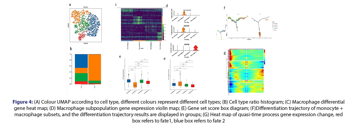
There were significant differences in the proportion of macrophage subgroups before and after treatment. After treatment, MAC-2 was the main component, while the proportion of other types of macrophages decreased significantly (Figure 4A,B). Different subsets of macrophages have different functional characteristics (Figure 4C,D), cluster 2, the significantly differentially expressed gene SELENOP is a characteristic gene of macrophages in recent studies.
Figure 4: (A) Colour UMAP according to cell type, different colours represent different cell types; (B) Cell type ratio histogram; (C) Macrophage differential gene heat map; (D) Macrophage subpopulation gene expression violin map; (E) Gene set score box diagram; (F)Differentiation trajectory of monocyte + macrophage subsets, and the differentiation trajectory results are displayed in groups; (G) Heat map of quasi-time process gene expression change, red box refers to fate1, blue box refers to fate 2
Locus analysis of monocytes and 4 macrophage subsets was performed to describe the differentiation and development direction of monocytes and macrophages in the samples. The differentiation of MAC-2 in pre-treatment samples was similar to that of Extracellular Matrix (ECM) macrophages (Figure 4E-G). The MAC-2 cells in the treated tissues were at the very end of differentiation. The results indicated that MAC cells could be induced to historesident differentiation after treatment.
Crosstalk of microenvironment before EC treatment
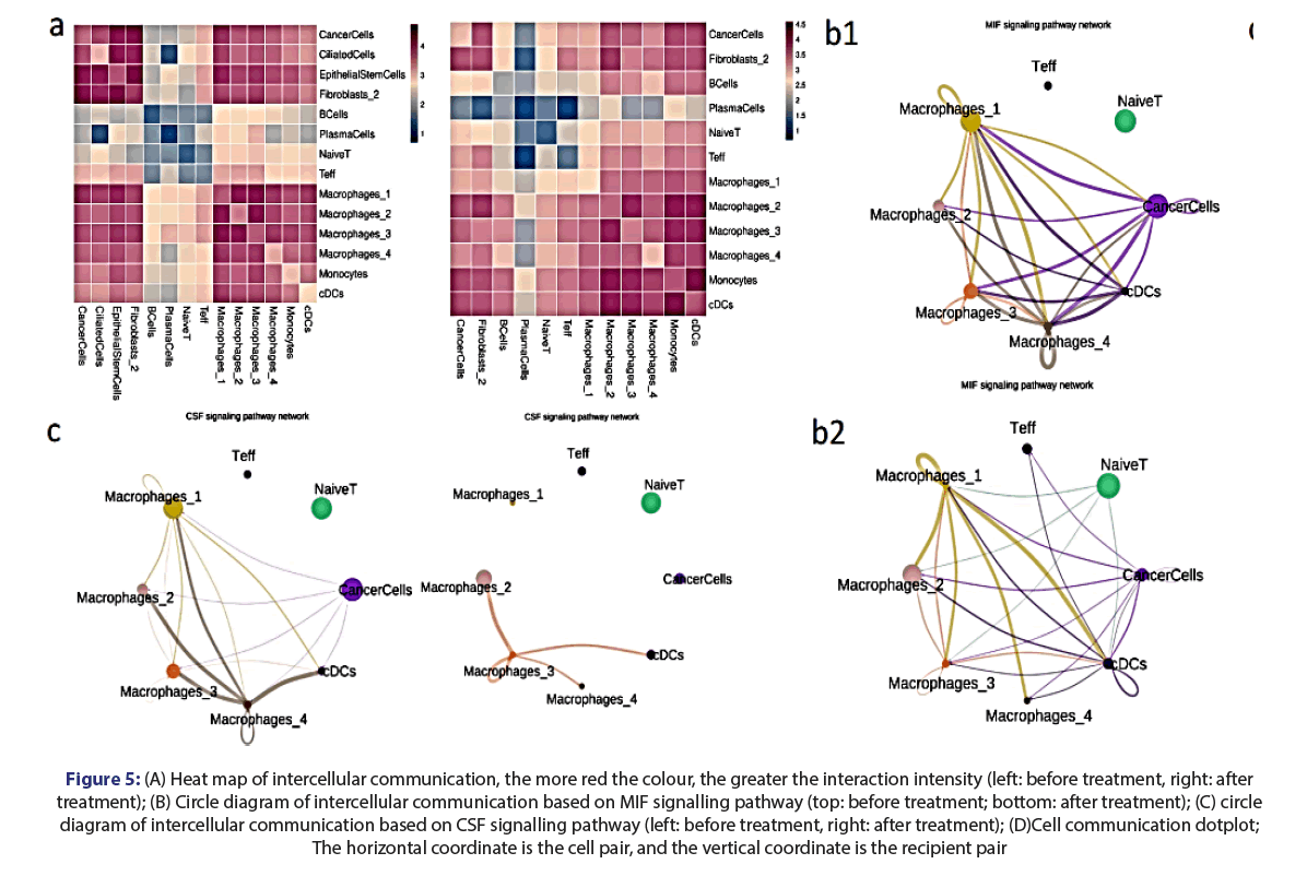
Based on the statistical analysis of cell interaction, it was found that the communication between macrophage subsets and DC cells in the samples and naiveT and Effector T Cell (TEFF) increased significantly after treatment compared with before and after treatment (Figure 5A). The communication between myeloid and lymphoid immune cells and tumor cells also increased after treatment. The APC in the samples after treatment was more focused on the antigen presenting ability of T cells (Figure 5B). In this study, intercellular (macrophage and tumor cell) communication based on Colony Stimulating Factors (CSF) signaling pathway was significantly reduced after hapten treatment, suggesting that it can be involved in regulating macrophage function after treatment (Figure 5C). The regulatory pathway between immune cells and tumor cells is learned through analysis based on signaling pathways, and the ligand pairs are further analyzed (Figure 5D).
Figure 5: (A) Heat map of intercellular communication, the more red the colour, the greater the interaction intensity (left: before treatment, right: after treatment); (B) Circle diagram of intercellular communication based on MIF signalling pathway (top: before treatment; bottom: after treatment); (C) circle diagram of intercellular communication based on CSF signalling pathway (left: before treatment, right: after treatment); (D)Cell communication dotplot; The horizontal coordinate is the cell pair, and the vertical coordinate is the recipient pair
The relationship between tumor gene expression and treatment
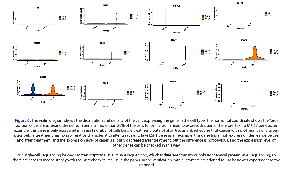
The violin diagram shows the distribution and density of the cells expressing the gene in the cell type (Figure 6). The horizontal coordinate shows the “proportion of cells” expressing the gene. In general, more than 25% of the cells to form a violin need to express this gene. Therefore, taking MKI67 gene as an example, this gene is only expressed in a small number of cells before treatment, but not after treatment, reflecting that cancer with proliferative characteristics before treatment has no proliferative characteristics after treatment. Taking (Estrogen Receptor Alpha) ESR1 gene as an example, this gene has a high expression profile before and after treatment, and the expression level of caner is slightly decreased after treatment, but the difference is not obvious. Other gene expression levels can be checked in this way.
Figure 6: The violin diagram shows the distribution and density of the cells expressing the gene in the cell type. The horizontal coordinate shows the “proportion
of cells” expressing the gene. In general, more than 25% of the cells to form a violin need to express this gene. Therefore, taking MKI67 gene as an example, this gene is only expressed in a small number of cells before treatment, but not after treatment, reflecting that cancer with proliferative characteristics
before treatment has no proliferative characteristics after treatment. Take ESR1 gene as an example, this gene has a high expression demeanor before and after treatment, and the expression level of caner is slightly decreased after treatment, but the difference is not obvious, and the expression level of other genes can be checked in this way
Ps: Single-cell sequencing belongs to transcriptome level mRNA sequencing, which is different from immunohistochemical protein level sequencing, so there are cases of inconsistency with the histochemical results in the paper. In the verification part, customers are advised to use basic wet experiment as the standard
Discussion
In the current study, we provide evidence that hapten enhanced intratumoral chemotherapy treatment of endometrial cancer induced acute immune response followed the hysterectomy, which, in turn, help to control the metastasis and recovery of cancer cells.
Our study demonstrate that the proportion of stromal cells increased and the number of tumor cells was significantly reduced after treatment, indicating the possible effectiveness of the treatment, the proportion of cells in the immune microenvironment was explained by subdivision annotation. The results of GSVA analysis showed enrichment after treatment: non-classical NF-kB activation, and the non-classical NF-κB pathway was responsible for the development of multilayer immune cells. This pathway is necessary for the maturation and function of TEC (Thymus Epithelial Cells), which is essential for T cell development in the thymus [16]. This was also consistent with an increase in Teff after treatment. Enrichment of endometrial cancer specificity in HALLMARK_PANCREAS_BETA_CELLS suggested that this could be a target for drug action, however, it is necessary to further verify the abnormal activation of enriched HEDGEHOG signaling pathway before treatment through β-catenin to promote endometrial cancer [17]. The activation of PI3K/AKT/mTOR signaling pathway mediates tumorigenesis, and the complex promotes EC progression through this pathway [18]. The evolutionally-conserved Notch signaling pathway regulates various cellular processes, such as proliferation, differentiation, and cell invasion. There is growing evidence linking abnormal Notch signaling to diseases such as hyperplasia and endometrial cancer [19].
Our study also showed the T cell function in immune activation, during the process of differentiation, there were high expression of T cell toxicity (CCL3, CLL4, GZMB, GZLY) and inflammation (CCL5, NGK7) genes in the intermediate stage. At the end of differentiation, T cells overexpress Kruppel Like Factor 6 (KLF6) and other genes: Among them, KLF6 is a known suppressor of various tumors [20].
FOS, FOSB, DUSP2 and GADD45B are genes related to inflammation and stress [21]. EGR1 induces T-BET transcription, suggesting a role in T cell activation [22]. Jun-modified CAR T cells can effectively improve the efficacy of T cells, alleviate cell failure and greatly enhance their ability to continuously clear tumors [23].
It was found that the proportion of cDCs increased and DC cells increased as the main APC cells in the microenvironment after treatment, suggesting that the antigen presenting ability of DC cells also increased after treatment, promoting T cells to kill tumors. Dendritic cells expressed high levels of B7-1 (CD80) and B7-2 (CD86) co-stimulatory factors, which fully activated T-fine inter-group gene expression [24]. The significant enrichment of MHC-related pathways in DC cells also suggests that T cell activation is driven by MHC signals provided by DC cells.
It was found that the macrophage increased after treatment as important factor in the microenvironment. MAC-2 was the main component, while the proportion of other types of macrophages decreased significantly. Different subsets of macrophages have different functional characteristics, cluster 2, the significantly deferentially expressed gene SELENOP is a characteristic gene of macrophages in recent studies, and has been shown to have anti-tumor effects in studies of non-small cell lung cancer [25]. Significantly high expression of CEBPD has been shown to induce apoptosis of cancer cells [26]. In the overall trajectory analysis diagram, three MAC-134 subgroups with ECM characteristics and MAC-2 with tissue resident characteristics were located in two opposite directions in the monocyte branch respectively.
It was found that the intercellular communication in MHC I pathway of concern showed that APC and T cells in MHC I pathway had obvious communication before and after treatment. The probability of communication between APC cells and T cell subsets increased after treatment. MHC Class I molecules mainly mediate the antigen presentation process of endogenous antigens, including tumor antigens synthesized in tumor cells and viral proteins synthesized by virus-infected cells [25].
In this study, it was found that the communication between macrophages and tumor cells based on MIF signaling pathway was also significantly reduced after treatment. LTB-LTBR and TNFSF14-LTBR are lymphocyte suppressor/activator pairs [26], targeting ltb-ltbr interaction can inhibit tumor growth and prolong the survival of colorectal cancer xenotransplantation patients [27]. Both immune cells and tumor cells had LTBR signaling interactions in the samples before and after treatment, indicating that these pathways are essential for the immune response against tumors. MIF is a widely expressed multipotent cytokine, which promotes tumor angiogenesis through recruitment of macrophages in colorectal cancer and further promotes tumor progression [28]. Moreover, MIF is increased in some tumors through chaperone stabilization of HSP90-related molecules [28]. After treatment, the expression of MIF-TNSFSF14 decreased, and the specific expression level decreased, indicating that the MIF signal was also decreased. The effect of CD74-MIF in Mac-4|caner interaction was significantly decreased after treatment versus before treatment, suggesting that the antigen could inhibit tumor inflammation and progression by inhibiting the action of macrophage migration factor. The clincalcopathological immunohistochemical results after treatment were consistent with the expression of these genes, which were examined by the technique scRNA-seq.
Conclusion
The relationship between tumor gene expression and treatment
The UMIPIC resulted in significantly clinical benefit, one local therapy with hapten and chemotherapy drugs can kill local endometrial cancer also induce immune response to fight cancer with or without hysterectomy and control expression of several genes related with endometrial cancer.
Strengths and limitations for treat endometrial cancer with HEIC
The results presented in our current study have never been demonstrated earlier by single therapy can induce immune response like immunotherapy and control expression of genes related endometrial cancer. A significant limitation of the study is the sample size: samples from only on patient were analyzed. Given the significant cost associated with scRNAseq, we will seek additional funding support to extend our study to more patient samples.
Our study provides evidence that hapten mediated local chemotherapy is safe and effective method while it induces a systematic immunity against cancer by initiating immune response from the endometrial cancer to achieve desirable clinical outcome in order to prevention of tumor cell metastasis with or without hysterectomy.
Ethical statement
All procedures and protocols in the study has been reviewed and approved by the Ethical Committee of the Beijing Baofa Cancer Hospital (TMBF 0010, 2015). All informed consent forms form patients have been signed prior to the start of the study.
References
- Amant F, Moerman P, Neven P, et al. Endometrial cancer. Lancet. 2005;366: 491-505.
- Trojano G, Olivieri C, Tinelli R et al. Conservative treatment in early stage endometrial cancer: a review. Acta Biomed. 2019;90:405-10.
- Aoki Y, Kanao H, Wang X, et al. Adjuvant treatment of endometrial cancer today. Jpn J Clin Oncol. 2020;50: 753-65.
- Jiménez JAM, Mulero SG, Matías-Guiu X et al. Facts and hopes in immunotherapy of endometrial cancer. Clin Cancer Res. 2022;28:4849-60.
- Gao F, Jing P, Liu J, et al.. Hapten-enhanced overall survival time in advanced hepatocellular carcinoma by ultro-minimum incision personalized intratumoral chemoimmunotherapy. J Hepatocell Carcinoma 2015;2: 57-68.
- Yu BF, HanY, Fu Q, et al. Awaken immune cells by hapten enhanced intratumoral chemotherapy with penicillin prolong pancreatic cancer survival. J Cancer. 2023; 14: 1282-92. [Crossref]
- Yu BF, Fu Q, HanY, et al. An acute inflammation with special expression of cd11 & cd4 produces abscopal effect by intratumoral injection chemotherapy drug with hapten in animal model. J Immunological Sci. 2022;6: 1-9.
- Ziegenhain C, Vieth B, Parekh S, et al. Comparative analysis of single-cell RNA sequencing methods. Mol Cell 2017;65: 631-43.
- Qing Liu S, Jie Gao Z, Wu J, et al. Single-cell and spatially resolved analysis uncovers cell heterogeneity of breast cancer. J Hematol Oncol. 2022;15:19.
- Kechin A, Boyarskikh U, Kel A, et al. cutPrimers: A new tool for accurate cutting of primers from reads of targeted next generation sequencing. J Comput Biol, 2017;24: 1138-43.
- Chen Z, Zhao M, Liang J, et al. Dissecting the single-cell transcriptome network underlying esophagus non-malignant tissues and esophageal squamous cell carcinoma. EBioMedicine, 2021;69: 103459.
- Dobin A, Davis CA, Schlesingeret F, et al. STAR: ultrafast universal RNA-seq aligner. Bioinformatics, 2013;29:15-21.
- Yu G, Wang LG, Han Y, et al. clusterProfiler: an R package for comparing biological themes among gene clusters. Omics, 2012;16: 284-7.
- Efremova M, Vento-Tormo M, Teichmann SA, et al. CellPhoneDB:inferring cell-cell communication from combined expression of multi-subunit ligand-receptor complexes. Nat Protoc. 2020;15:1484-506.
- Qiu X, Hill A, Packeret J, et al. Single-cell mRNA quantification and differential analysis with Census. Nat Methods, 2017;14: 309-15.
- Yu H, Lin L, Zhang Z, et al. Targeting NF-κB pathway for the therapy of diseases: mechanism and clinical study. Signal Transduct Target Ther. 2020;5: 209.
- Liao XY, Siu MKY, Au CWH, et al. Aberrant activation of hedgehog signaling pathway contributes to endometrial carcinogenesis through beta-catenin. Mod Pathol. 2009;22: 839-47.
- Liao J, Chen H, Qi M, et al. MLLT11-TRIL complex promotes the progression of endometrial cancer through PI3K/AKT/mTOR signaling pathway. Cancer Biol Ther. 2022;23: 211-24.
- Jonusiene V and Sasnauskiene A. Notch and Endometrial Cancer. Adv Exp Med Biol. 2021;1287:47-57.
- Slavin DA, Koritschoner NP, Prieto CC, et al. A new role for the Kruppel-like transcription factor KLF6 as an inhibitor of c-Jun proto-oncoprotein function. Oncogene. 2004;23: 8196-205.
- Ye Y, Chen Z, Jiang S, et al. Single-cell profiling reveals distinct adaptive immune hallmarks in MDA5+ dermatomyositis with therapeutic implications. Nat Commun. 2022;13:6458.
- Shin HJ, Lee JB, Park SH, et al. T-bet expression is regulated by EGR1-mediated signaling in activated T cells. Clin Immunol. 2009;131:385-94.
- Lynn RC, Weber EW, Sotillo E, et al. c-Jun overexpression in CAR T cells induces exhaustion resistance. Nature. 2019;576: 293-300.
- Tao Y, Li X, Yushan Zhang Y, et al. TP53-related signature for predicting prognosis and tumor microenvironment characteristics in bladder cancer: A multi-omics study. Front Genet. 2022;13: 1057302.
- Wang C, Qiuxiao Yu Q, Song T, et al. The heterogeneous immune landscape between lung adenocarcinoma and squamous carcinoma revealed by single-cell RNA sequencing. Signal Transduct Target Ther. 2022;7:289.
- Pan Y, Li C, Ko C, et al. CEBPD reverses RB/E2F1-mediated gene repression and participates in HMDB-induced apoptosis of cancer cells. Clin Cancer Res. 2010;16:5770-80.
- Pittet MJ, Michielin O, Denis Migliorini D. Clinical relevance of tumour-associated macrophages. Nat Rev Clin Oncol. 2022;19: 402-21.
- Juliane W, Abisola AO, Kevin MJ, et al. Concepts of extracellular matrix remodelling in tumour progression and metastasis. 2020;11:5120.


